| Source | M.W. | 38 - 41 kDa | CAS No. | ||
|---|---|---|---|---|---|
| Structural Info | |||||
| Formulation | Lyophilized in sterile filtered solution of PBS with 1% CHAPS | ||||
| Reconstitution | Before reconstitution, we recommend a brief spin to drive down any material dislodged from the bottom of the tube. The lyophilized protein should be reconstituted in sterile H2O to a concentration of 100 ng/uL. Because of the hydrophobic nature of this protein, further dilutions should be made in buffer or medium containing carrier proteins, such as albumin or serum. | ||||
| Stability | The lyophilized protein is stable for at least 6 months if stored at -80 °C. Reconstituted protein is stable for at least two weeks at 4 °C, but should be stored in aliquots at -80 °C for longer term. Avoid repeated freeze and thaw. |
||||
| Purity | Greater than 90% as determined by SDS-PAGE and HPLC analysis | ||||
| Biological Activity | The activity was determined by using a TCF reporter gene assay in cultured human cells. The EC50 ranges from 50 - 150 ng/ml. | ||||
| Country of Origin | USA | ||||
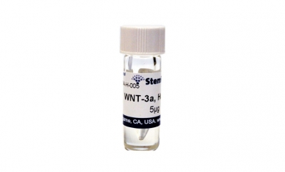
WNT-3a is a member of the WNT family of signaling proteins that play key roles in embryonic development and the maintenance of adult tissues. WNT-3a is a prototypic canonical WNT that signals through the b-catenin pathway. The predicted size of human WNT-3a is a monomeric protein containing 328 amino acid residues. Due to glycosylation, it migrates at an apparent molecular weight of 38 - 41 kDa on SDS-PAGE under non-reducing conditions. StemRD’s WNT-3a is produced from a human cell line overexpressing human Wnt-3a cDNA in protein-free medium. Purification is done by a proprietary process that is distinct from the published method.
Kim SE, Huang H, Zhao M, et al., Wnt Stabilization of β-Catenin Reveals Principles for Morphogen Receptor-Scaffold
Assemblies. Science. 2013 May 17; 340(6134):867-70
http://www.ncbi.nlm.nih.gov/pubmed/23579495
Zhang X, Abreu JG, Yokota C, et al., Tiki1 is required for head formation via Wnt cleavage-oxidation and inactivation. Cell. 2012 Jun 22;149(7):1565-77.
http://www.ncbi.nlm.nih.gov/pubmed/22726442
Hoshiba T, Kawazoe N, Chen G. Mechanism of regulation of PPARG expression of mesenchymal stem cells by osteogenesis-mimickingextracellular matrices. Biosci Biotechnol Biochem. 2011;75(11):2099-104.
http://www.ncbi.nlm.nih.gov/pubmed/22056426
